

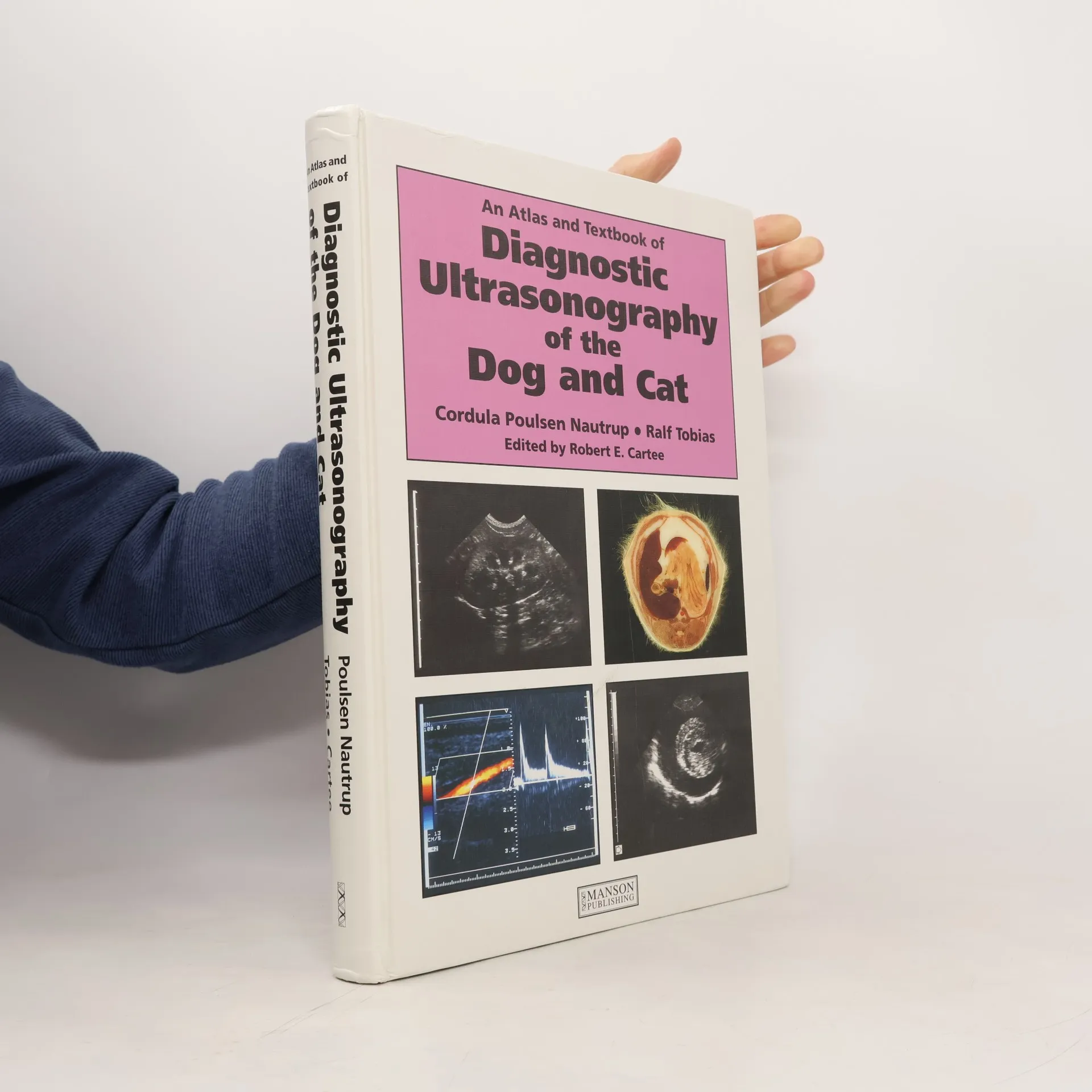


An Atlas and Textbook of Diagnostic Ultrasonography of the Dog and Cat
Viac o knihe
Veterinarians need increasingly to be familiar with the interpretation of ultrasound imaging. In this magnificent volume, the authors set standards as they describe all the main aspects of sonography affecting diagnosis in dogs and cats, including ultrasound, m-mode, pulsed and color Doppler echography. Particular attention is devoted to echocardiography, abdominal and pelvic sonography, and foetal ultrasonography. This book also deals with recognition of the disease process and courses of treatment. The authors further illustrate cases by using conventional radiography, computed microfocal tomographs, photographs of specimens and line drawings. Designed and produced for ease of use this volume has become a source of constant and essential reference for veterinarians in training, practice, teaching, and research that need to interpret ultrasound imaging.
Nákup knihy
An Atlas and Textbook of Diagnostic Ultrasonography of the Dog and Cat, Cordula Poulsen Nautrup, Ralf Tobias, Robert E. Cartee
- Jazyk

- Rok vydania
- 2000
- Väzba
- (pevná)
Doručenie
Platobné metódy
Nikto zatiaľ neohodnotil.
- Titul
- An Atlas and Textbook of Diagnostic Ultrasonography of the Dog and Cat
- Jazyk
- anglicky
- Vydavateľ
- Manson Publishing
- Rok vydania
- 2000
- Väzba
- pevná
- Počet strán
- 400
- ISBN10
- 1874545103
- ISBN13
- 9781874545101
- Série
- Štítky
- Medicína, Odborná literatúra, Psy, Mačky, Choroby, Atlasy, Veterinárne lekárstvo
- Anotácia
- Veterinarians need increasingly to be familiar with the interpretation of ultrasound imaging. In this magnificent volume, the authors set standards as they describe all the main aspects of sonography affecting diagnosis in dogs and cats, including ultrasound, m-mode, pulsed and color Doppler echography. Particular attention is devoted to echocardiography, abdominal and pelvic sonography, and foetal ultrasonography. This book also deals with recognition of the disease process and courses of treatment. The authors further illustrate cases by using conventional radiography, computed microfocal tomographs, photographs of specimens and line drawings. Designed and produced for ease of use this volume has become a source of constant and essential reference for veterinarians in training, practice, teaching, and research that need to interpret ultrasound imaging.